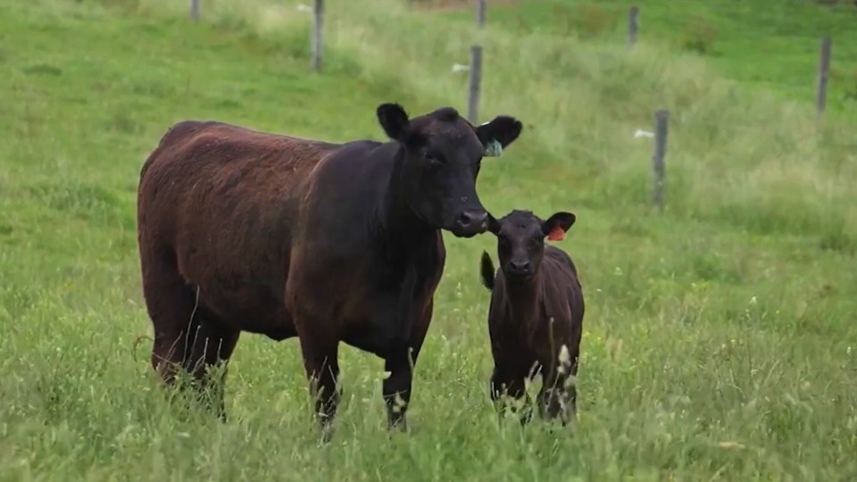
Keep bovine coronavirus (BCV) on your radar
By Dr. Brent Meyer
When I think about bovine infectious diseases, especially bovine respiratory and enteric diseases, bovine coronavirus (BCV) speeds to the front. I often investigate both scour and respiratory outbreaks in beef/dairy herds, and a common finding with diagnostics is BCV.
Given the frequent occurrence of BCV — and the significant impact it can have on the growth and productivity of beef cattle — producers should be aware of the presentation, prevention, and treatment of the virus and consider discussing it with their veterinarian.
BCV is well documented for its role in enteric disease and until recently, was considered solely an enteric pathogen. It affects the small intestines and colon and is especially lethal in young calves due to the rapid loss of bicarbonate in feces. BCV is the cause of winter dysentery in adult cows. However, growing evidence shows its prevalence and association with respiratory disease.1

BCV has an affinity to both intestinal and respiratory cellular structures. BCV destroys goblet cells, which line both the trachea and the intestinal tract. These are the cells that produce a “mucus mat,” which is the calf’s defense against invasion of pathogenic bacteria.
We often see a combination of BCV with Mannheimia haemolytica, Pasteurella multocida, Histophilus somni and Mycoplasma bovis. If these bacteria are present with a concurrent BCV infection, then the bacteria have an unobstructed path into the lungs of a calf to develop bacterial pneumonia. In comparison to the enteric form, it is well established that BCV can be lethal by itself, but often allows for colonization with Rotavirus, Escherichia coli or Salmonella sp.
BCV enteric presentation is typically seen in young calves (3-21 days old) and is characterized by profuse watery or hemorrhagic diarrhea, with high morbidity and mortality rates. From a respiratory standpoint, it often is seen just prior to turnout or branding, preweaning and during the first 45 days in the feed yard.
Uncomplicated cases of BCV generate a mild fever around 103 F. There can be clear nasal discharge and ocular discharge as well. Another common presentation is increased respiration rate and coughing.
Research shows that cattle shedding BCV from the nasal cavity and seronegative to BCV were 1.6 times more likely to require treatment for respiratory disease than cattle that did not shed virus or seropositive to BCV.2 Cattle shedding BCV were 2.2 times more likely to have pulmonary lesions at slaughter than cattle that did not shed BCV.
These findings support the theory that neutralizing antibodies in naturally exposed calves, or cattle arriving at feedlots, have been correlated to protection against respiratory disease, pneumonia, or BCV shedding.
Global prevalence
Results from ongoing and retrospective studies using 1363 nasal swabs collected from cattle experiencing BRD where one or more respiratory pathogens were detected (34% of samples) showed that BCV was detected most frequently (22.9%) compared to 11.6%, 7.0%, 6.1%, and 5.0% for BRSV, PI3, BHV-1, and BVDV, respectively.1,2-3
The virus presence has been confirmed on all continents, and seroprevalence studies demonstrate that over 90% of cattle have been exposed to BCV during their lifetime.4 Prevalence may be underestimated because uncomplicated respiratory BCV infection is often undiagnosed (samples not submitted) because it seldom results in extreme illness and self-cures within a week.
However, if calves progress to severe pneumonia, or non-responsive diarrhea, then nasal swabs or fecal samples should be submitted to a diagnostic laboratory.
To better protect animals, producers should know the common signs of BCV as well as practical management steps needed to decrease BCV-related illness in calves.
Vaccination and colostrum management
Currently, there are no vaccines available for the respiratory form of BCV. There are BCV vaccines available that are labeled for the enteric form. More research is needed on the application and effectiveness of respiratory BCV vaccines.
The number one management goal to help prevent BCV calf scours is ensuring that calves consume high quality colostrum from BCV-vaccinated dams. Calves should be up and nursing within six hours of birth, because by 12 hours, the esophageal groove that directs colostrum into the small intestine for absorption starts to seal up.
Administer a BCV-containing vaccine in cows and heifers prior to calving. This boosts antibody levels in the colostrum, providing newborn calves with increased protection. Adhere to the correct protocol for the vaccine and avoid administering the vaccine less than five weeks prior to calving. It takes about five weeks for the mammary gland to generate colostral antibodies, so administering a vaccine too close to calving may reduce its effectiveness.
The calving interval, labor and facilities determine the cow vaccination window. Choose a vaccine with an extended vaccination window before calving so you can guarantee colostrum quality. It’s also important to choose a broad-spectrum vaccine with antigens associated with major bacterial and viral causes of scours in young calves, including Rotavirus, Coronavirus types 1 & 3, Clostridium perfringens types C & D and Escherichia coli type K99.
If a producer happens to miss the cow vaccination protocol, then there are a few vaccines labeled for BCV enteric protection in the neonatal calf. In neonatal calves, intranasal BCV vaccines are effective against enteric BCV infections and avoid interference from maternal antibodies.
Consult with your veterinarian to determine the appropriate vaccine and vaccination schedule for your herd.
Environmental management
The most common way that BCV spreads is through direct contact between infected and susceptible cattle and fecal-oral transmission. BCV can also live on surfaces for a long time if not inactivated by soaps or disinfectants.
To decrease the incidence of BCV illness in calves, spread out your calving groups and rotate/clean areas to avoid BCV spread. Utilizing a Sandhills or Modified Sandhills calving system greatly reduces enteric issues in calves. Contaminated feed and water sources can act as potential sources of infection. Clean and disinfect all equipment (bottles, esophageal feeders, boots and coveralls).
Do not commingle calves from other farms or your own calves from different locations or calving groups. Implement efforts to minimize the stress of transportation and abrupt changes in the environment.
Treatment of BCV
Early detection, proper supportive care, and veterinary intervention help lessen the effects of BCV while minimizing complications, alleviating symptoms and promoting faster recovery.
Treatment for enteric BCV is heavily dependent on oral and/or IV fluids. Excessive loss of fluids and electrolytes from prolonged and severe diarrhea can disrupt the balance within the body, leading to dehydration. Dehydration can further exacerbate the animal’s condition and increase the risk of complications. Oral fluids alone are often not enough for severe BCV, so IV fluids for two to three days in the clinic or on the ranch may be recommended.
Treatment for respiratory BCV is dependent on what respiratory bacteria is of concern. Antibiotics have no effect on virus treatment, but they do minimize the secondary bacterial infections from making the situation worse.
Regularly monitor the progress of infected animals and follow up with your veterinarian to assess the effectiveness of treatment. Adjustments to the treatment plan may be necessary based on the individual animal’s response.
Bovine coronavirus poses a significant challenge to beef cattle herds, impacting animal health and profitability. Always consider submitting samples to the diagnostic lab if BCV is on your radar. Understanding how to prevent and manage BCV through comprehensive management approaches will lower the incidence of BCV-caused illness and lessen the impact it has on the health of your herd.
References
- PM Hick, AJ Read, I. Lugton, F. Busfield, KE Dawood, L. Gabor, M. Hornitzky and PD Kirkland. Coronavirus Infection in Intensively Managed Cattle with Respiratory Disease. Australian Vet. J. 2012. 90.10: 381-386.
- Oystein Angen, John Thomsen, Lars Erik Larsen, Jesper Larsen, Branko Kokotovic, Peter M H Heegaard and Jorg M D Enemark. Respiratory Disease in Calves: Microbiological Investigations on Trans-tracheally Aspirated Bronchoalveolar Fluid and Acute Phase Protein Response. Vet. Microbiology. 2009. 137: 165-171.
- O’Neill, R., J. Mooney, E. Connaghan, C. Furphy and D.A. Graham. Patterns of Detection of Respiratory Viruses in Nasal Swabs from Calves in Ireland: A Retrospective Study. Vet. Record. July 21, 2014. Tech. no. 10.1136/vr.102574. veterinaryrecord.bmj.com.
- Vlasova, A. N. and Saif, L. J. Bovine Coronavirus and the Associated Diseases. Frontiers in Vet. Sci. 2021. 8, 643220. https://doi.org/10.3389/fvets.2021.643220.
Find more content for your beef operation.
About the author

Brent Meyer
D.V.M., M.S.,
Technical Services Veterinarian,
Merck Animal Health
